Table of Contents
- What Is Cryptorchid in Dogs?
- Health Risks of Cryptorchidism in Dogs
- Diagnosis: Physical Examination and Imaging
- Cryptorchid Neuter Cost and Surgical Options
Last Updated: July 12, 2026
What Is Cryptorchid in Dogs?
A cryptorchid is a congenital condition in which one or both testicles fail to descend into the scrotum. This guide from CorePet covers everything dog owners need to know about retained testicles, from diagnosis through surgical management. According to veterinary research, cryptorchidism affects approximately 1-10% of male dogs, making it one of the most common reproductive abnormalities in canine health. The condition can be unilateral (one testicle retained) or bilateral (both testicles undescended), and it requires veterinary intervention to prevent serious health complications.
The retained testicle typically remains in the inguinal canal or abdominal cavity along the path of normal descensus testis. Unlike a normal testicle that descends during fetal development, a cryptorchid testicle becomes trapped due to anatomical abnormalities in the gubernaculum or surrounding tissues. This distinction matters because an undescended testis functions differently than a properly descended one, creating unique risks for your pet.
Health Risks of Cryptorchidism in Dogs
The retained testicle creates several serious health concerns that extend beyond simple infertility. An undescended testis experiences higher internal body temperature, which damages the tissue responsible for spermatogenesis and leads to testicular atrophy over time. This temperature difference between the scrotum and the abdomen or inguinal canal is the primary reason cryptorchid testicles fail to function normally.
The most significant risk is testicular malignancy. Dogs with cryptorchidism have a substantially elevated risk of developing germ cell tumors, including seminoma and other cancers. Research indicates that retained testicles are 4-8 times more likely to develop malignant tumors compared to normally descended testicles. This risk increases with age, making early surgical intervention critical for long-term health.
Additional complications include testicular torsion, where the spermatic cord twists and cuts off blood supply to the retained testicle. This creates sudden, severe pain and requires emergency surgical removal. Infection and inflammation of the cryptorchid testicle can also occur, causing systemic illness and discomfort. The longer a testicle remains undescended, the greater the cumulative risk of these complications.
Diagnosis: Physical Examination and Imaging
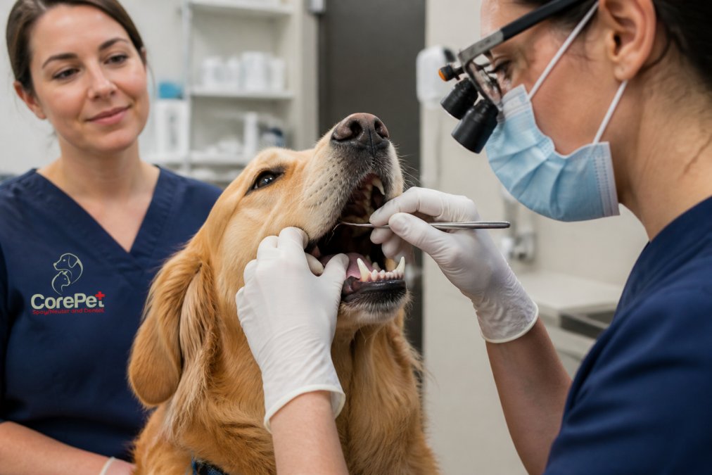
Diagnosing cryptorchidism begins with a thorough physical examination. A veterinarian will palpate the scrotum and inguinal canal to determine whether one or both testicles are absent from the scrotum. If one testicle cannot be felt in its normal location, the dog is classified as having a non-palpable cryptorchid testicle, which means the retained testicle is likely located in the abdominal cavity rather than the inguinal canal.
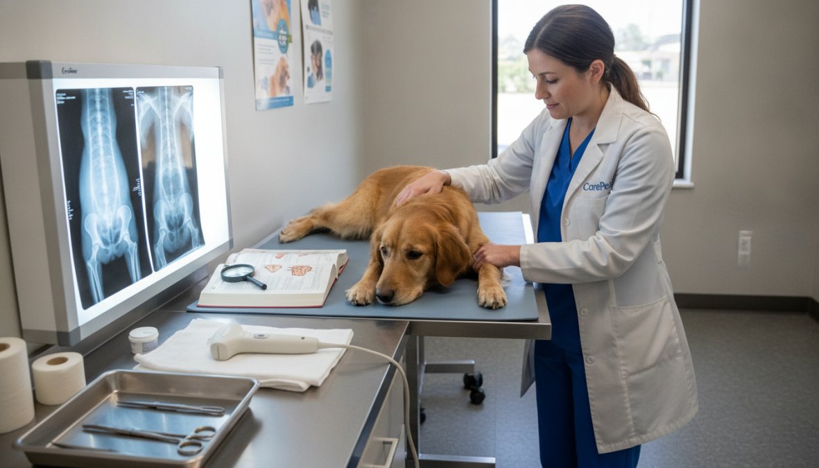
Ultrasonography is the primary imaging modality used to locate a non-palpable cryptorchid testicle. Ultrasound can visualize the retained testicle within the abdomen or inguinal canal with high accuracy, providing crucial information about size, shape, and tissue quality. The procedure is non-invasive, requires no anesthesia, and gives the veterinarian a clear picture of where the testicle is located before surgery.
For cases where ultrasonography is inconclusive or when a testicle cannot be located in the expected path, MRI or CT imaging may be recommended. These advanced imaging modalities provide detailed cross-sectional views of the abdomen and can identify a cryptorchid testicle in unusual locations. However, ultrasound is typically sufficient for surgical planning in most cases.
A palpable cryptorchid testicle, one that can be felt in the inguinal canal, is easier to diagnose and locate surgically. The veterinarian can often feel the retained testicle as a small, firm structure in the groin area. Even if palpable, imaging confirmation helps determine the best surgical approach and ensures no second cryptorchid testicle is missed during examination.
Cryptorchid Neuter Cost and Surgical Options
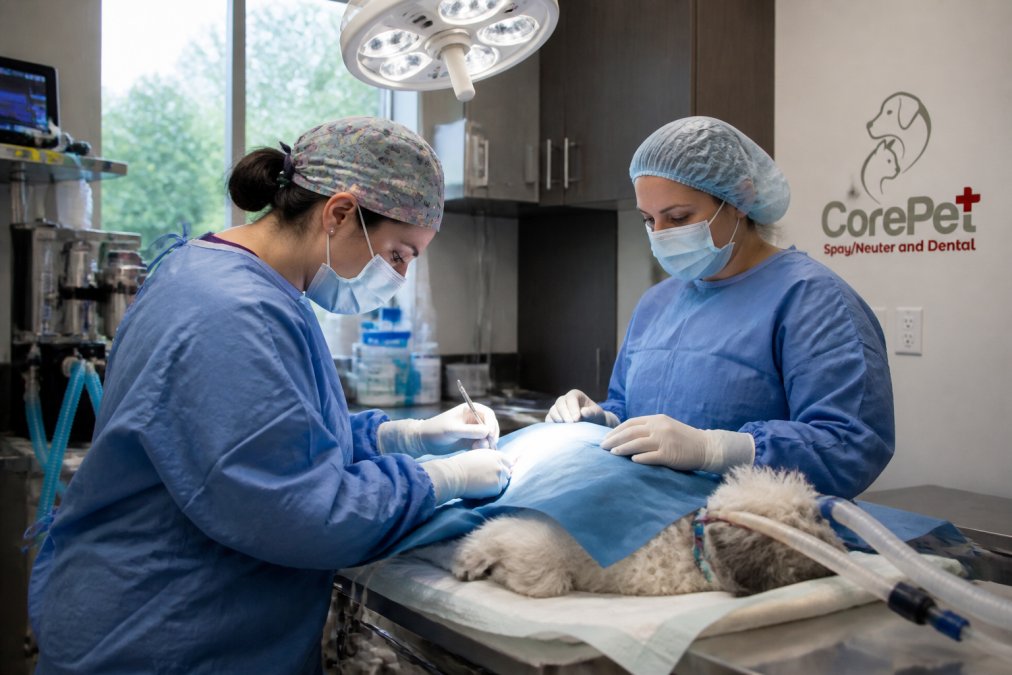
Cryptorchid neuter surgery differs from routine castration because the veterinarian must locate and remove a testicle that is not in its normal position. The complexity of the procedure depends on whether the retained testicle is palpable (in the inguinal canal) or non-palpable (in the abdomen). CorePet Surgical Center in Bedford, New Hampshire focuses on reproductive procedures at more affordable costs than many traditional veterinary hospitals, and Spay Neuter Dental services are available to address your dog’s surgical needs comprehensively.
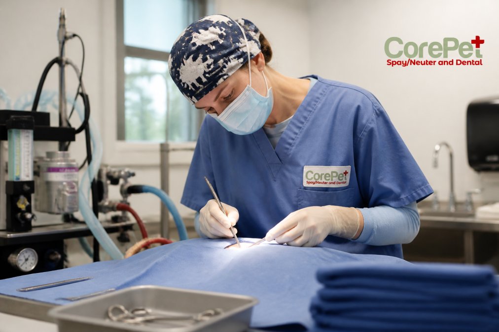
A palpable cryptorchid removal requires a small incision over the inguinal canal to access and extract the retained testicle. This procedure is straightforward and typically takes 20-30 minutes. The surgeon removes the cryptorchid testicle along with the normally descended testicle through separate incisions, completing the neuter in a single anesthetic event.
Non-palpable cryptorchid cases require abdominal surgery, which is more involved. The veterinarian makes an incision into the abdomen to locate the retained testicle along the path of descensus testis.
|
Surgical Approach |
Incision Size |
Recovery Time |
Anesthesia Duration |
|---|---|---|---|
|
Palpable inguinal removal |
Small (0.5-1 inch) |
7-10 days |
20-30 minutes |
|
Non-palpable abdominal surgery |
Larger (1-2 inches) |
10-14 days |
45-60 minutes |
Post-operative care for cryptorchid neuter involves restricting activity for 10-14 days, keeping the incision clean and dry, and administering prescribed pain medication as directed. Most dogs recover well from the procedure and show immediate improvement in comfort once the cryptorchid testicle is removed. The risk of future testicular malignancy is eliminated, and the dog can live a normal, healthy life.
Cryptorchidism is a manageable condition when addressed promptly with surgical intervention. CorePet Surgical Center in Bedford, New Hampshire focuses on reproductive procedures with advanced surgical techniques and modern equipment, ensuring safe, individualized care at reasonable costs. By choosing early neuter surgery for a cryptorchid dog, you eliminate the risk of testicular malignancy, prevent painful complications like testicular torsion, and give your dog the best chance at long-term health. Book an appointment with CorePet today to discuss your dog’s cryptorchid condition and explore the surgical options that work best for your pet’s needs.
Frequently Asked Questions
What does it mean if a dog is cryptorchid?
A cryptorchid dog has one or both testicles that fail to descend into the scrotum. Instead, the testicle remains in the abdomen or inguinal canal. This is a congenital condition where the gubernaculum—the structure that normally guides testicular descent—does not function properly. Cryptorchidism requires veterinary attention because retained testicles carry significant health risks.
What are the main health risks of cryptorchidism?
Retained testicles face dramatically increased risk of testicular malignancy, including seminoma and other germ cell tumors. They also experience compromised spermatogenesis (sperm production), leading to infertility. Additionally, undescended testicles are prone to testicular torsion, atrophy, and rupture. Early surgical intervention significantly reduces these risks and improves long-term health outcomes.
How is cryptorchid neuter surgery different from a standard neuter?
Standard neuter surgery removes both palpable testicles from the scrotum. Cryptorchid neuter requires locating a retained testicle in the abdomen or inguinal canal, often using ultrasonography or laparoscopy. Non-palpable testicles may necessitate exploratory abdominal surgery or minimally invasive laparoscopic techniques. CorePet Surgical Center focuses on these reproductive procedures at affordable costs with modern surgical techniques.
What is the recovery timeline after retained testicle surgery?
Most dogs recover within 10–14 days, though activity restrictions typically last 2–3 weeks. Post-operative care includes pain management, incision monitoring, and limiting running or jumping. Long-term quality of life is excellent when surgery is performed before malignant transformation occurs. Follow your veterinarian's aftercare instructions carefully to ensure proper healing and prevent complications.
This article was written using GrandRanker